 EVERYTHING


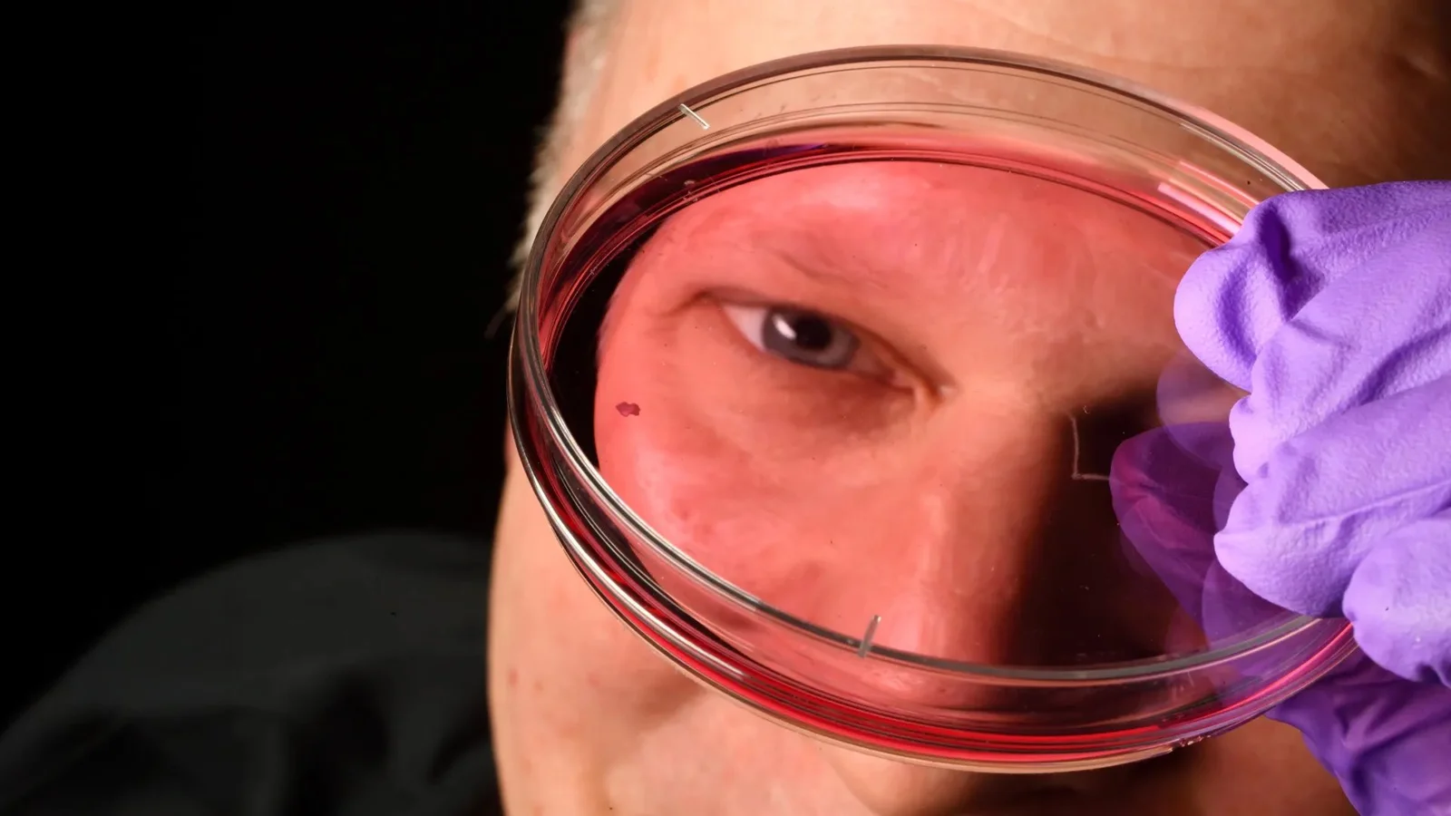
According to Sciencedaily, researchers at Johns Hopkins University have uncovered a surprising biological process that dictates how humans develop sharp central vision before birth. The study identifies a precisely timed interaction between thyroid hormones and molecules derived from vitamin A within the retina. This discovery is significant because it provides a clearer understanding of the foveola, the tiny region responsible for approximately half of all human visual perception.
Mechanisms of Retinal Development
The research team utilized retinal organoids—small clusters of tissue grown from fetal cells that mimic human retinal structures—to observe cellular changes over several months. They focused specifically on cone photoreceptors, which are responsible for daytime and color vision. While the rest of the retina contains a mix of blue, green, and red cones, the foveola is unique because it contains only red and green cones to facilitate high-acuity sight.
The study revealed that this specialized arrangement occurs through a two-step sequence during fetal development between weeks 10 and 14:
- Retinoic acid, a molecule derived from vitamin A, is broken down to limit the formation of new blue cones.
- Thyroid hormones then act on the remaining blue cone cells, driving them to convert into red and green cones.
- This coordinated transition ensures that blue cones do not persist in the foveola, which would otherwise impair visual clarity.
Challenging Existing Scientific Models
For decades, scientists struggled to explain this process because common laboratory models, such as mice and fish, do not develop the same photoreceptor arrangements as humans. The new findings suggest that rather than blue cones migrating away from the center of the retina, they undergo a physical transformation into other types.
"First, retinoic acid helps set the pattern. Then, thyroid hormone plays a role in converting the leftover cells," — Robert J. Johnston Jr., an associate professor of biology at Johns Hopkins who led the research.p>
Implications for Vision Restoration
Understanding these specific pathways is critical for addressing conditions like macular degeneration and glaucoma, which primarily damage the central retina. By mastering how these cells transform in a lab setting, scientists hope to eventually grow and transplant functional retinal tissues. This research provides a foundational step toward creating biologically accurate organoids that could one day restore sight to those with degenerative eye diseases.